Digital radiography
To assist the dentist in making diagnostic-therapeutic decisions with regard to biological and environmental effects, radiological imaging must be used and supported.
In the dental industry, X-rays are still frequently used for routine procedures. However, conventional x-rays required film processing. Dental offices had to physically store and distribute the copies, which was a time-consuming and expensive process.
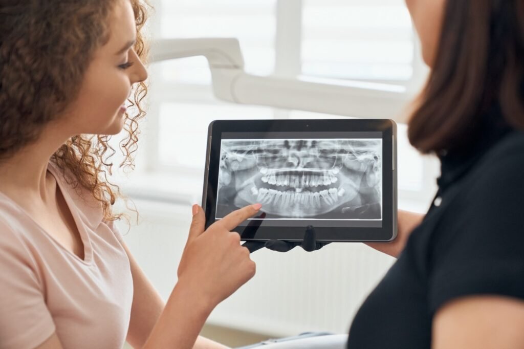
That has changed with direct digital radiography. The digital radiographic images produced by this sophisticated X-ray inspection technique appear on a computer screen quickly.
Now the most prevalent advanced dental technology used by patients during diagnostic visits is digital radiography.
Digital x-rays can be taken intraorally (inside the mouth) and extraorally (outside the mouth) by a dental professional. After being scanned, that file is then kept in a physical server or the cloud, making distribution much simpler.
This dental technology also has the advantage of shorter exposure durations, enhanced linearity, instantaneous feedback, and simplifying data storage.
In addition, when compared to traditional 3D radiological techniques, low-dose and ultra-low-dose cone beam computed tomography (C.B.C.T.) has a number of advantages. The main benefit is that the patient is exposed to less radiation, which is important for younger patients and those who need multiple scans.
References
Price, J.B. (2023). Digital Imaging. In Clinical Applications of Digital Dental Technology (eds R. Masri and C.F. Driscoll). https://doi.org/10.1002/9781119800613.ch1
Distefano, Salvatore, Maria Grazia Cannarozzo, Gianrico Spagnuolo, Marco Brady Bucci, and Roberto Lo Giudice. 2023. “The “Dedicated” C.B.C.T. in Dentistry” International Journal of Environmental Research and Public Health 20, no. 11: 5954. https://doi.org/10.3390/ijerph20115954


